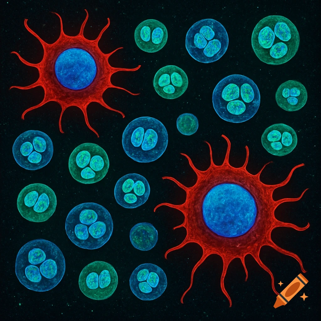
Immunofluorescence images of GAP-43 staining showing axonal regrowth post-tumor resection
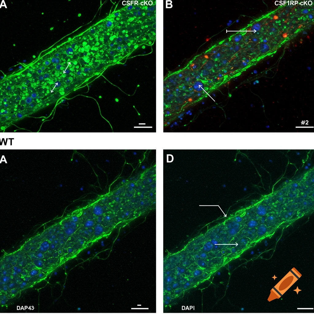
Microglial Activation and Axonal Regrowth Post-Tumor Resection (A) Immunofluorescence Image of GAP-43 Staining Description for AI Generator: Generate a side-by-side panel of two fluorescence microscopy images (one labeled "WT" and the other "CSF1R-cKO"). The images should show axonal regrowth in the peri-resection region, with GAP-43+ axons appearing in green and counterstained with DAPI (blue) for nuclei. In the WT image, depict numerous, long, regenerating axons, while in the CSF1R-cKO image, See more