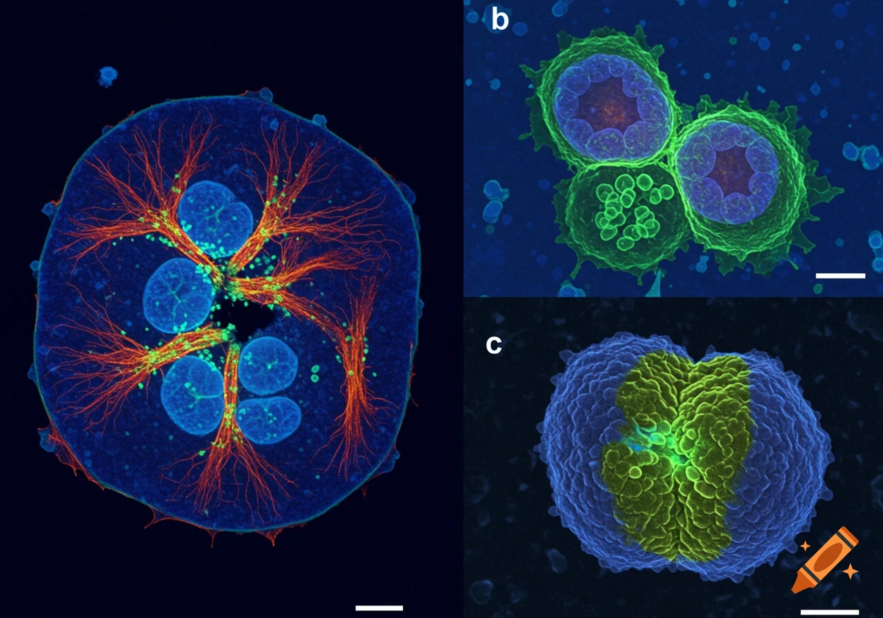
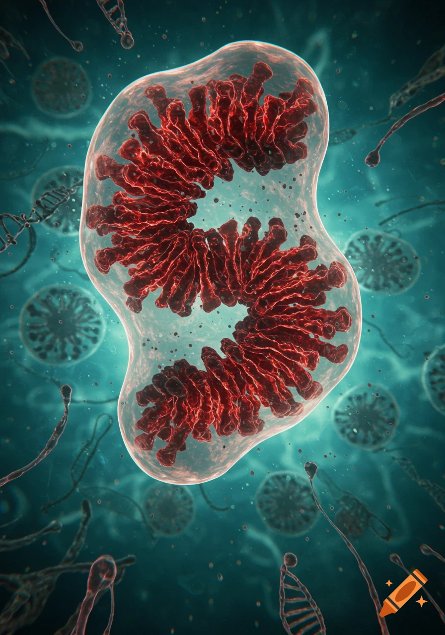
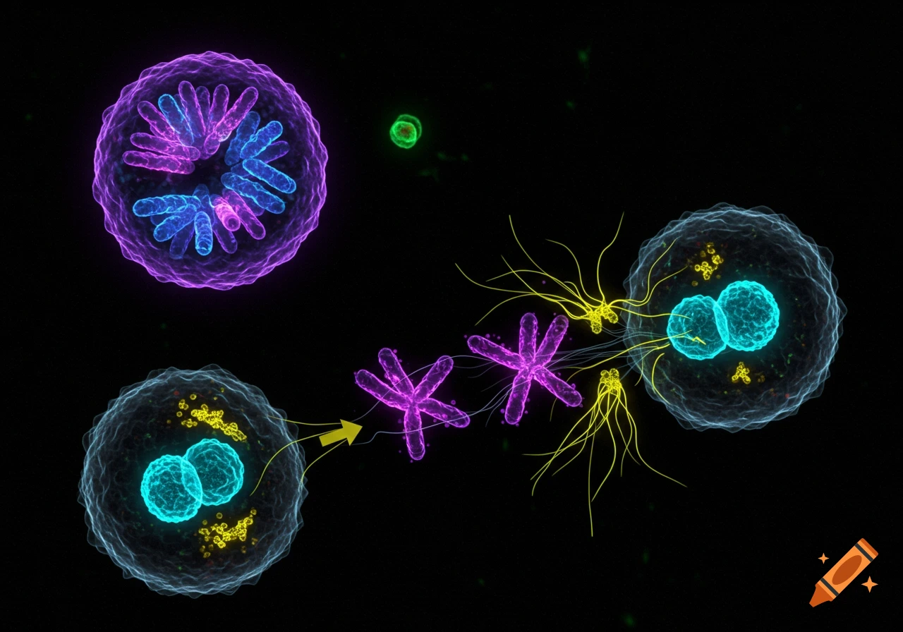
Fluorescent microscope image of cells with blue nuclei and red structures on a black background.
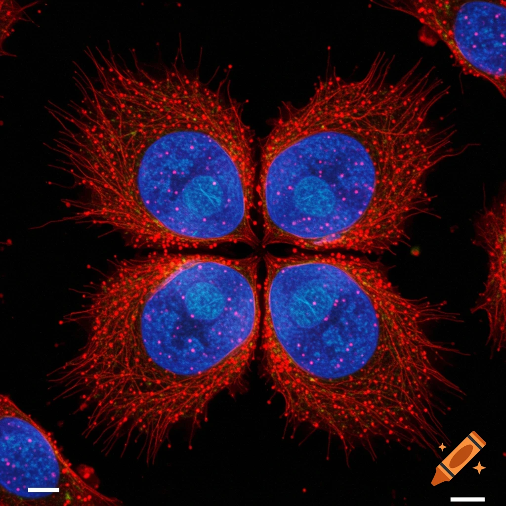
A mammalian cell. Three components have been fluoresced under a microscope. At the center of the cell, we have two blue oval nuclei mirroring one another back-to-back. The division occurred almost vertically, tilted 15 degrees to the right. We have red microtubules that are just the dots of spindles. Add anywhere from 25-50 of the red dots to the topmost portion of the nuclei shapes, the bottommost portion of the nuclei shapes, and then on each side of the groove of the two oval nuclei. The See more