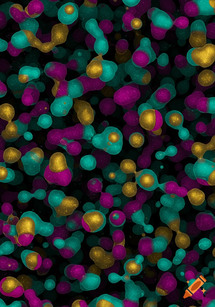
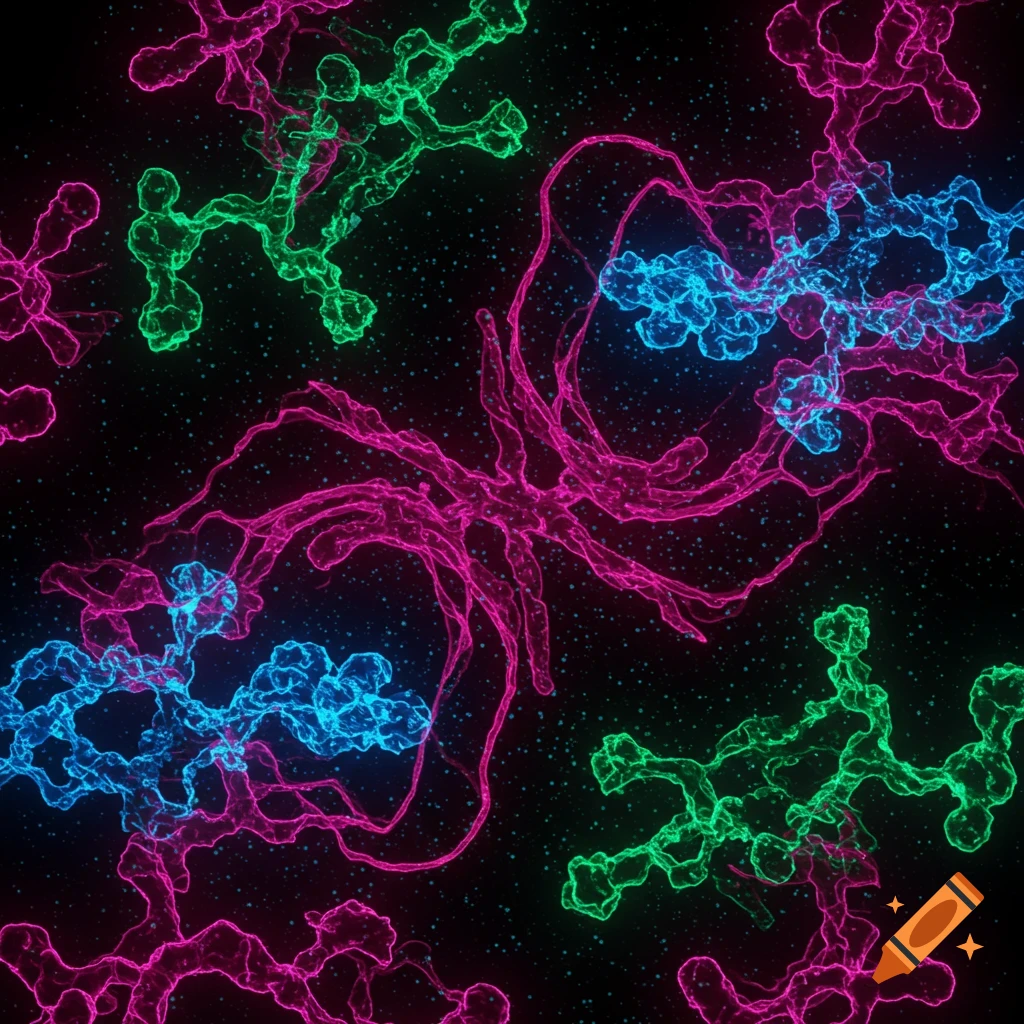
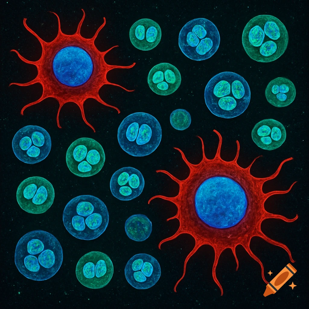
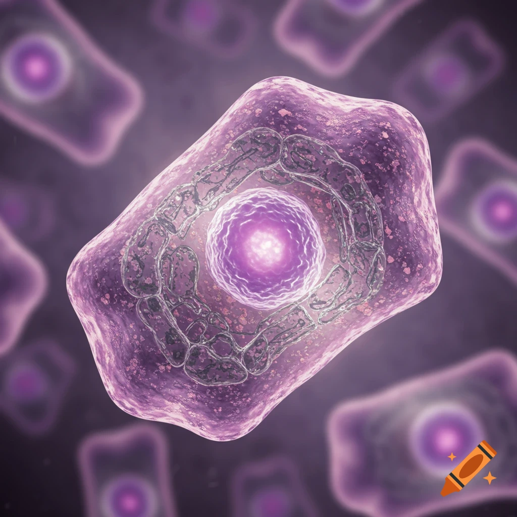
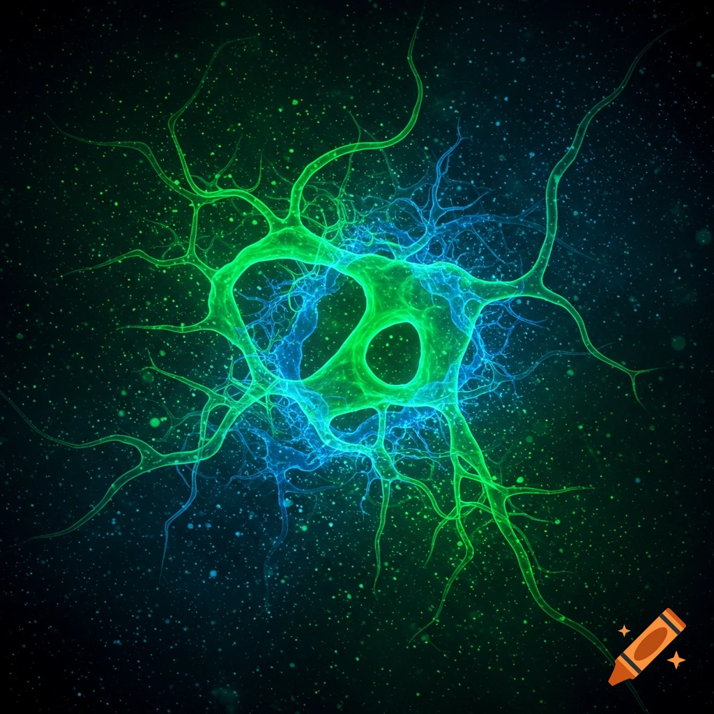
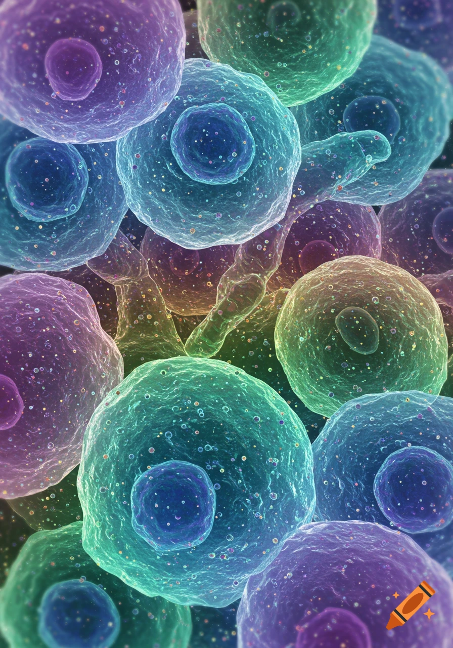
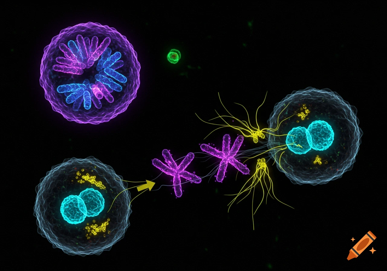
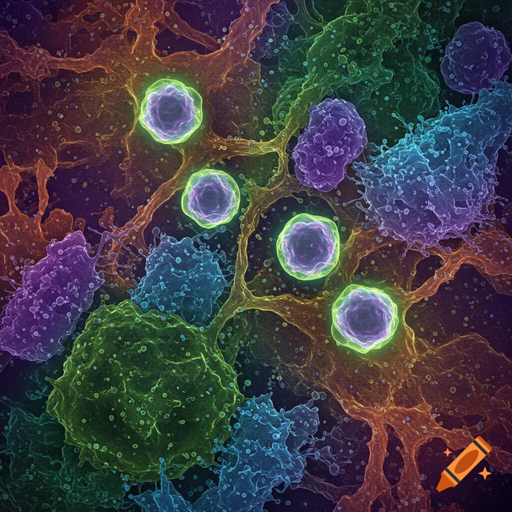
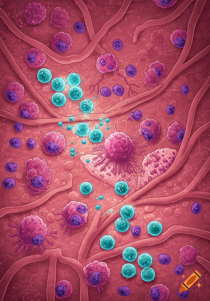
Fluorescence microscopy image showing purple cell nuclei surrounded by green cytoplasm on a dark background.
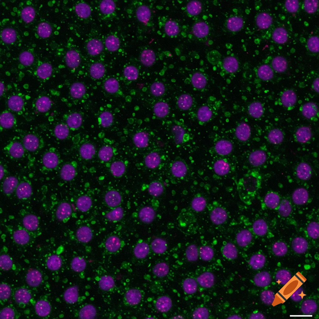
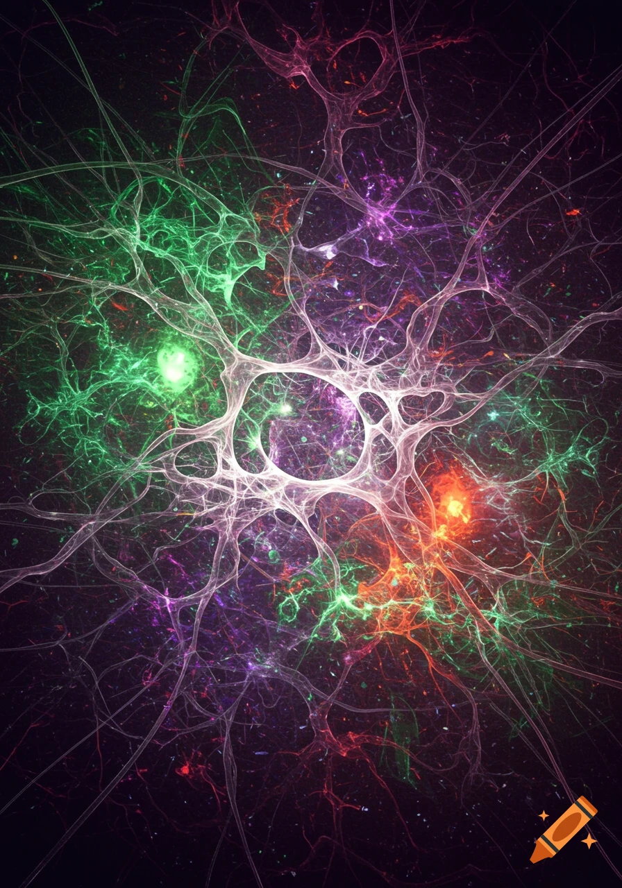
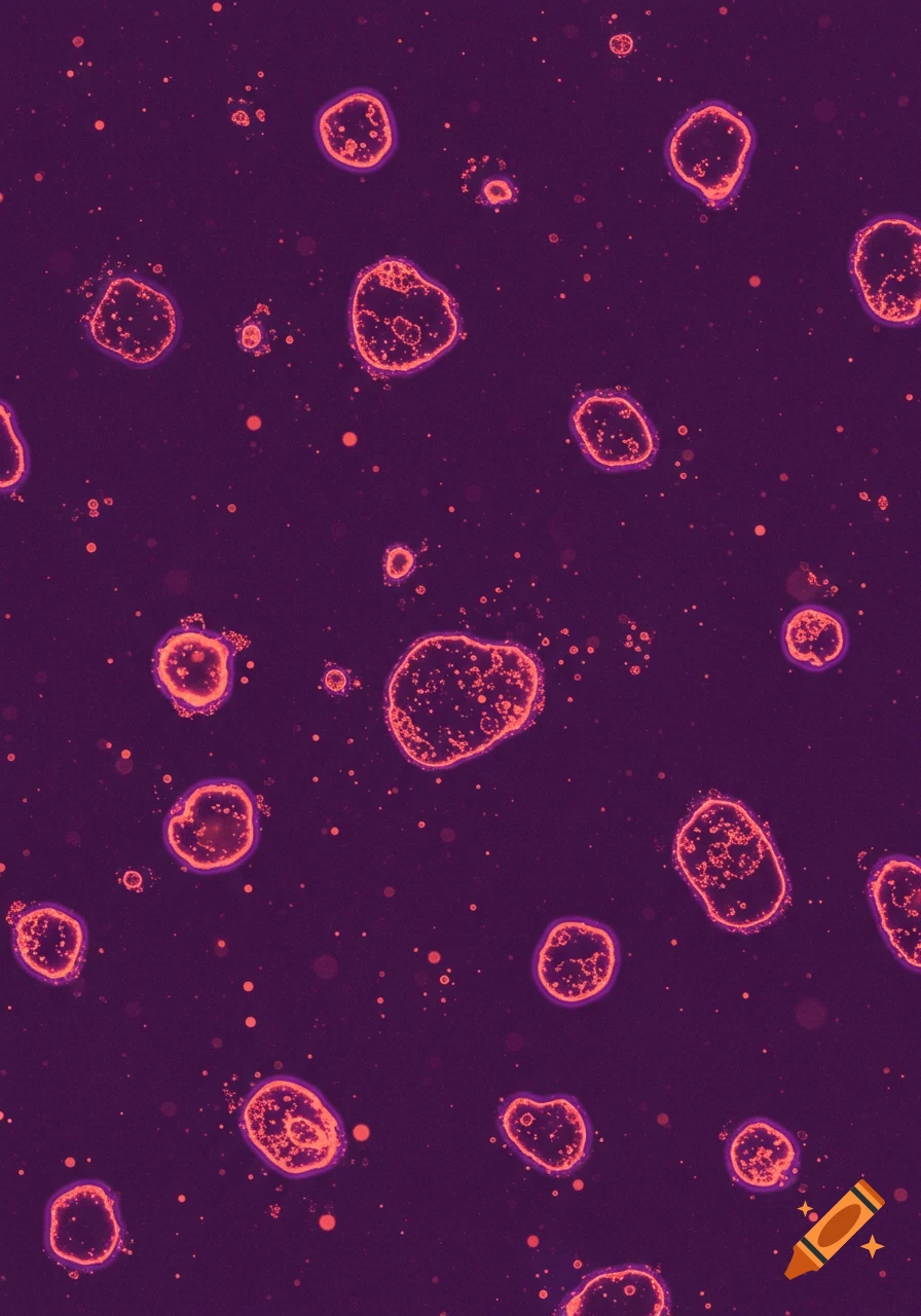
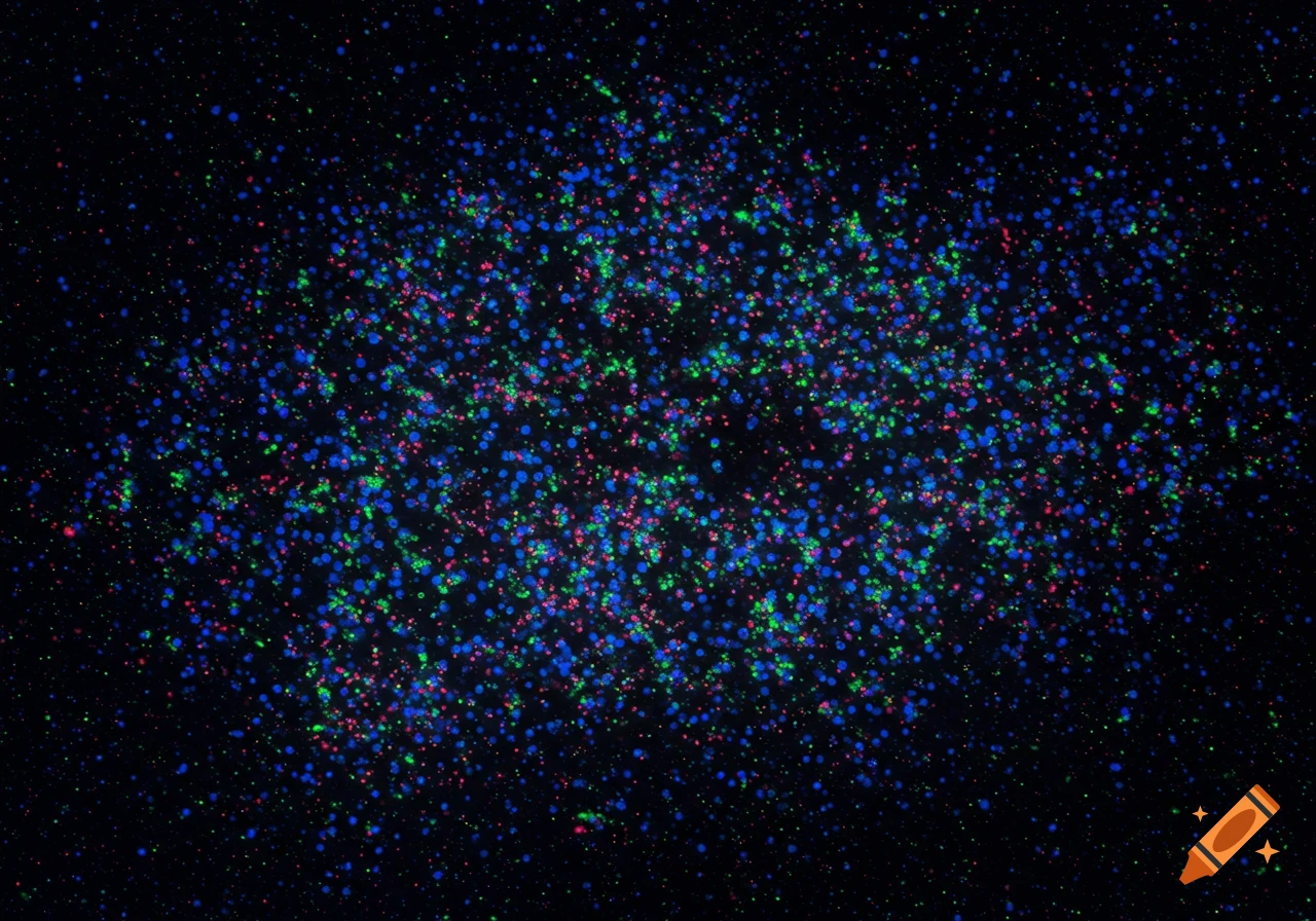
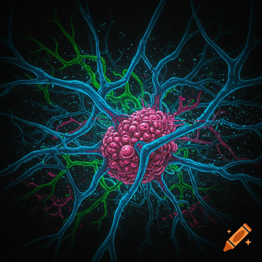
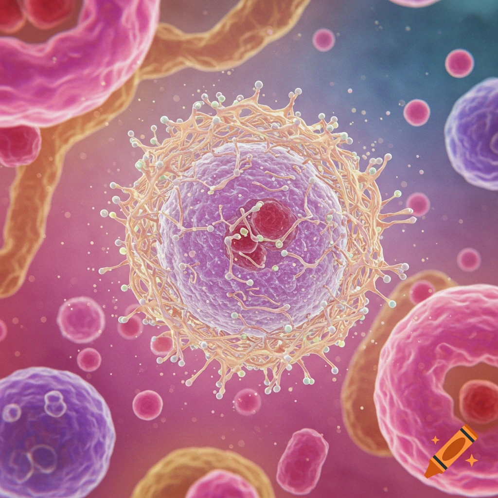
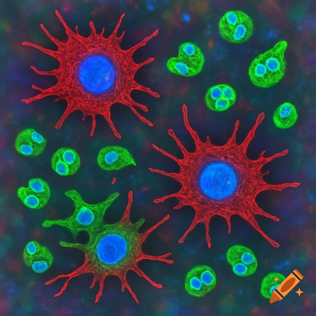
Fluorescence microscopy image of a mammalian brain or tissue sample. The background is dark green-black. Dozens of small, irregularly scattered cells appear, each with a glowing magenta nucleus (circular or oval) stained with DAPI or a similar nuclear dye. Surrounding many of these nuclei is green cytoplasmic or membrane staining (like Iba1 or microglial marker), appearing speckled or halo-like. Green does not completely fill the cells, but creates a cloudy, dispersed pattern around some See more