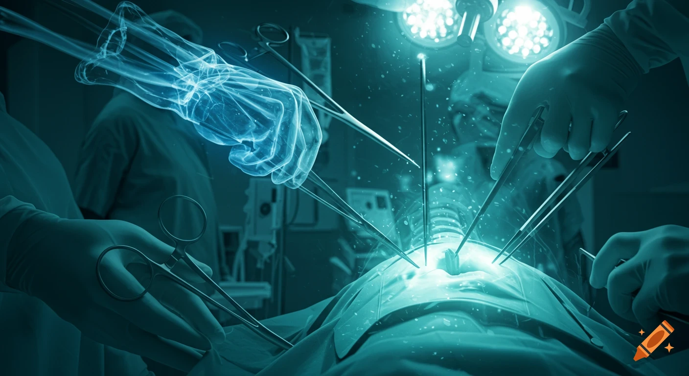
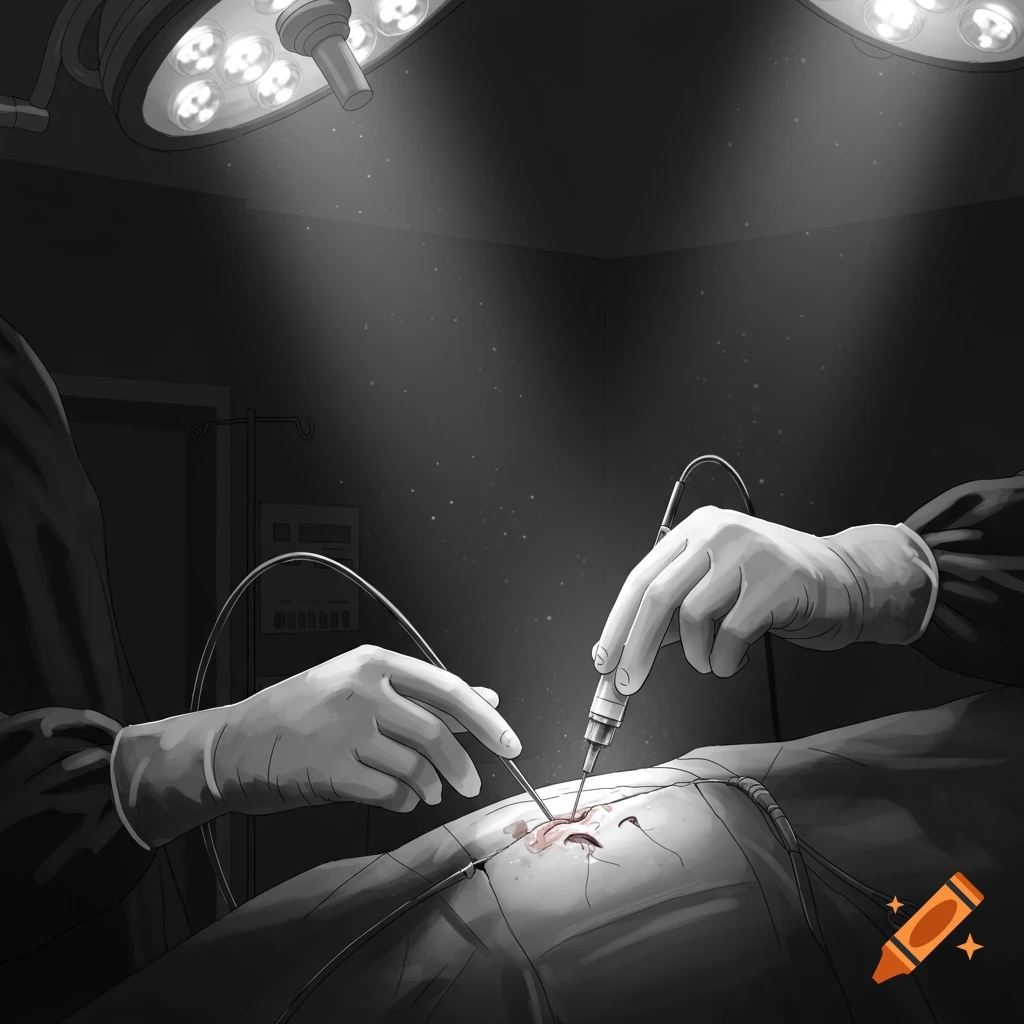
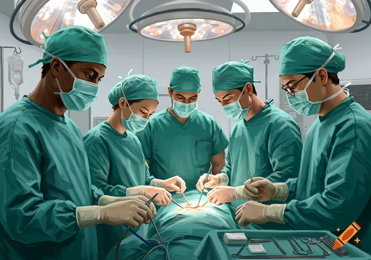
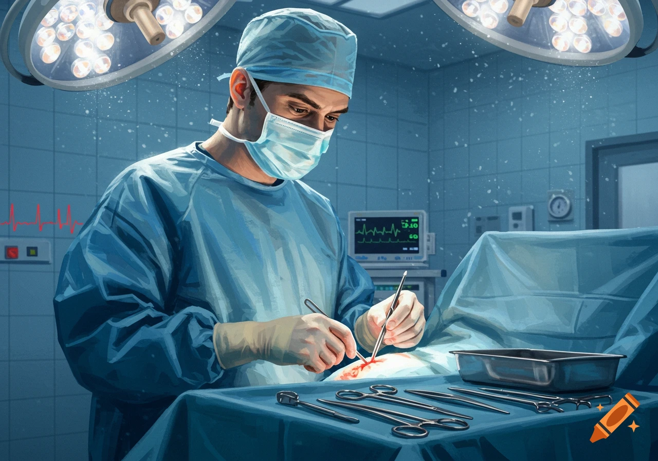
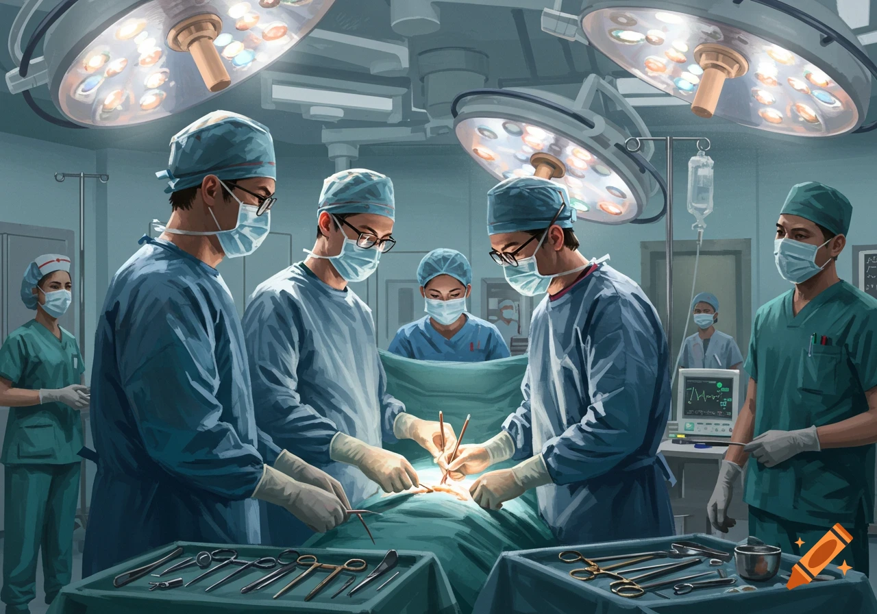
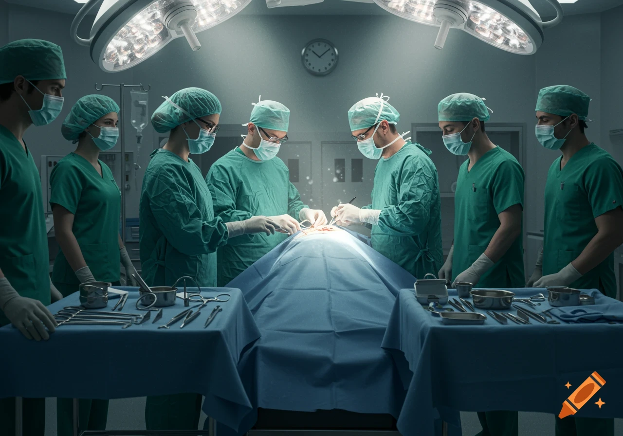
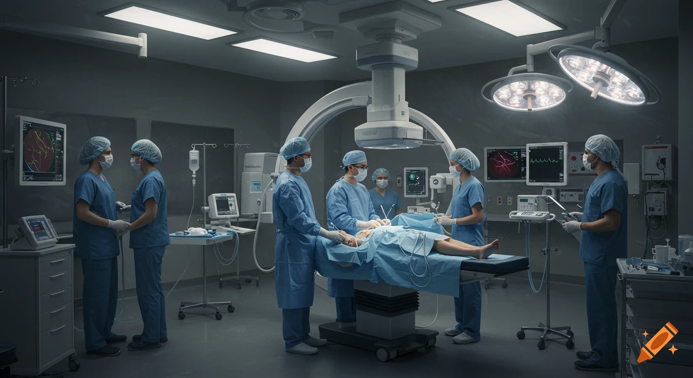
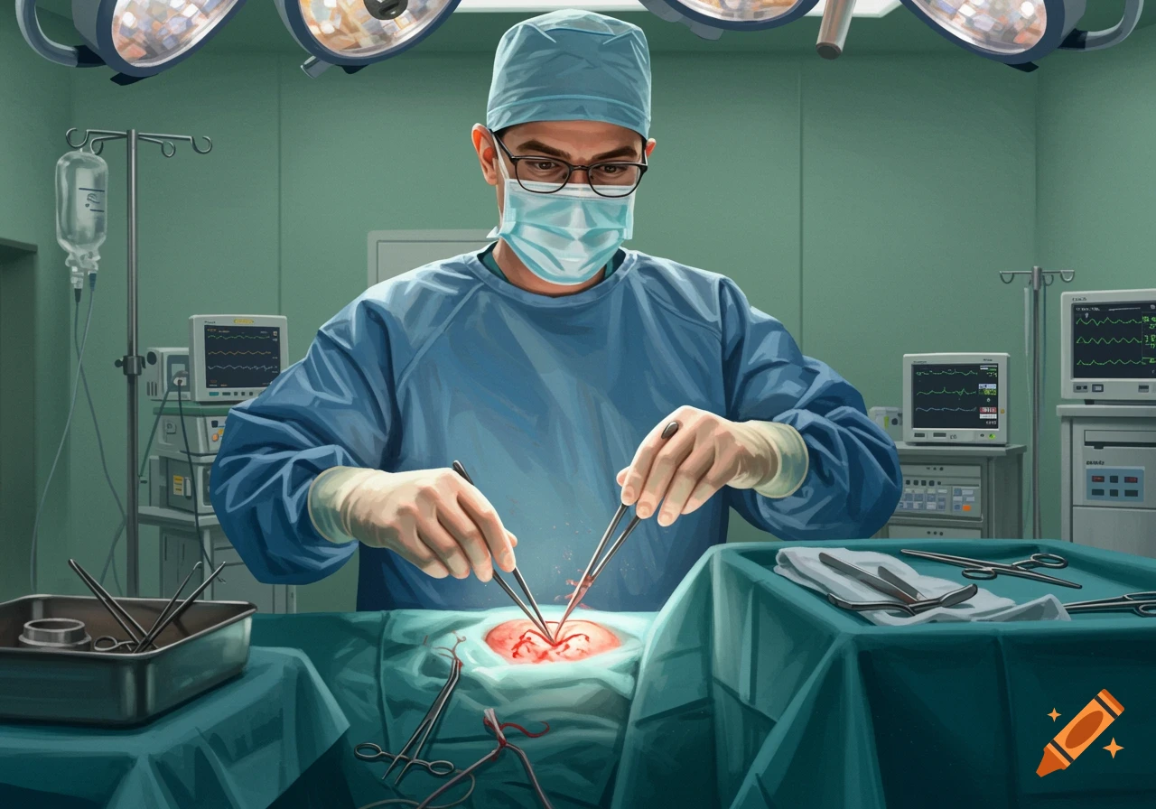
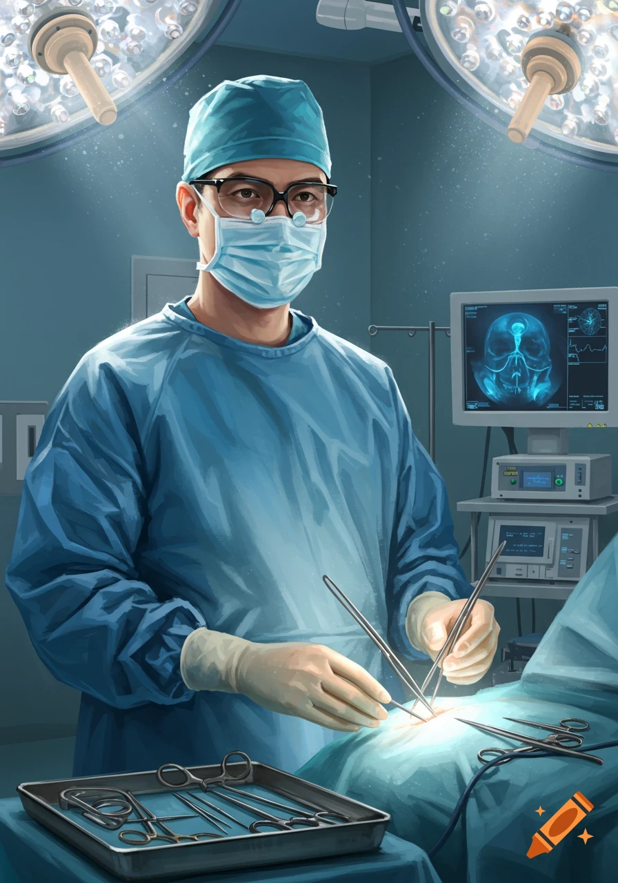
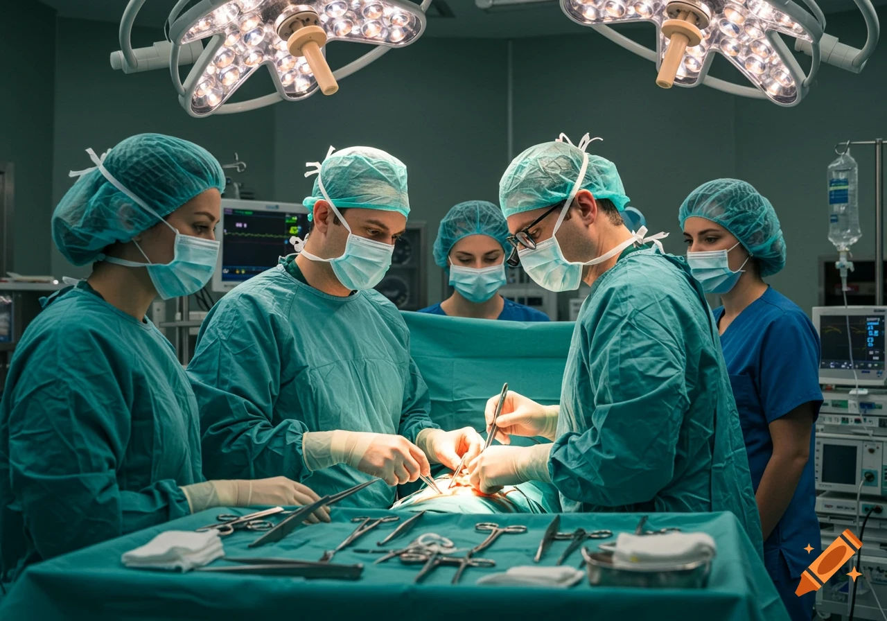
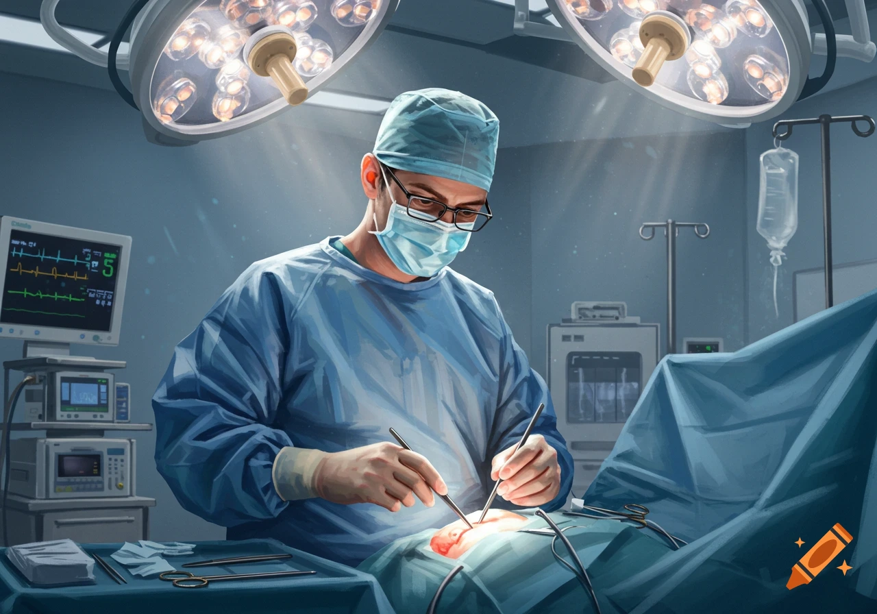
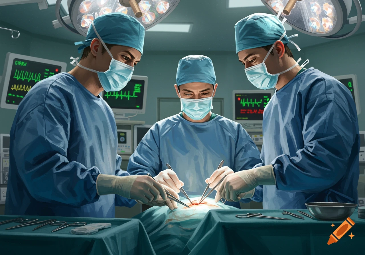
Detailed illustration of a surgical field under a microscope with various tools.
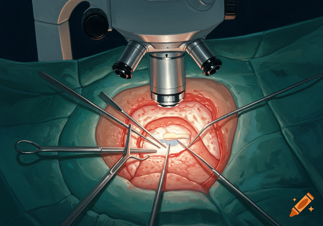
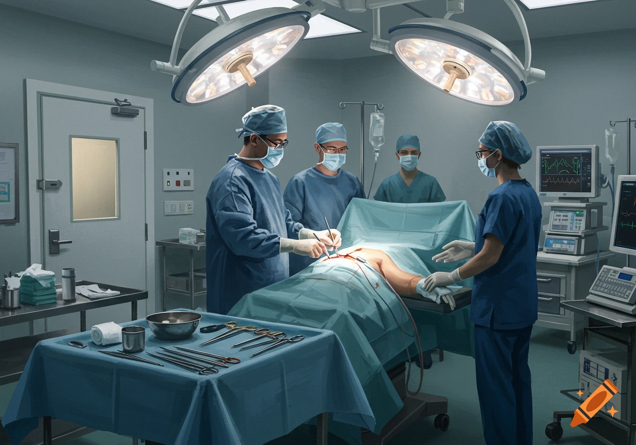
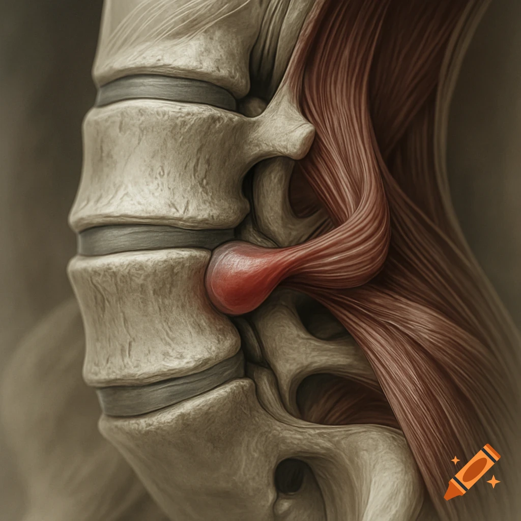
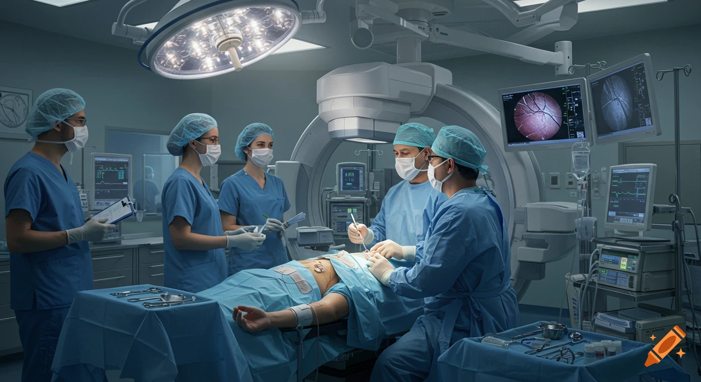
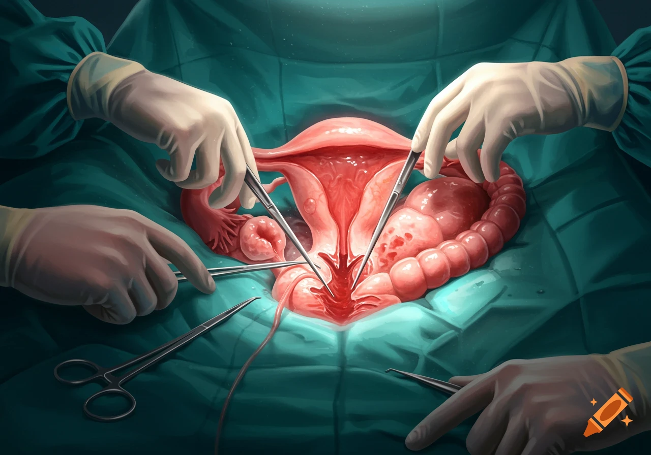
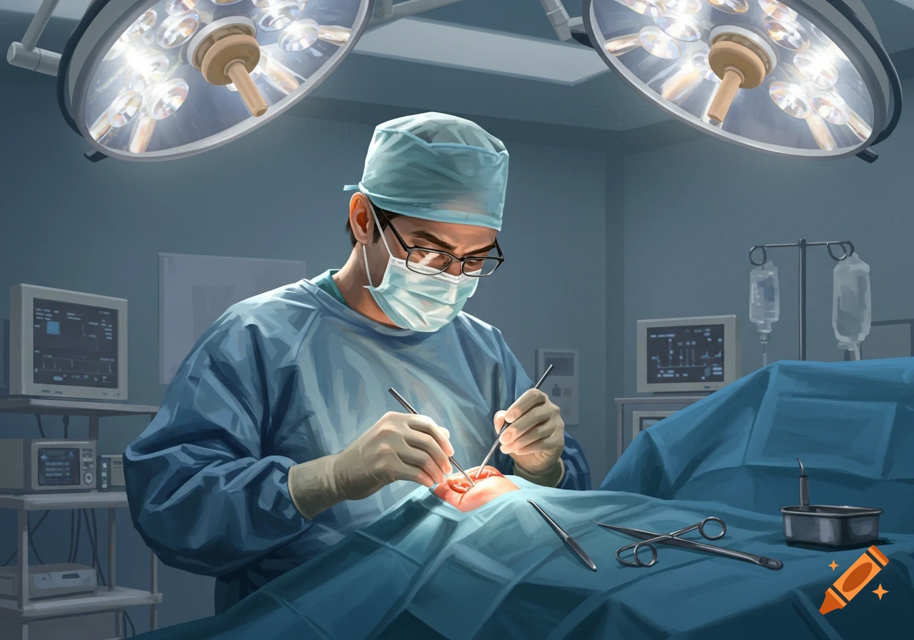
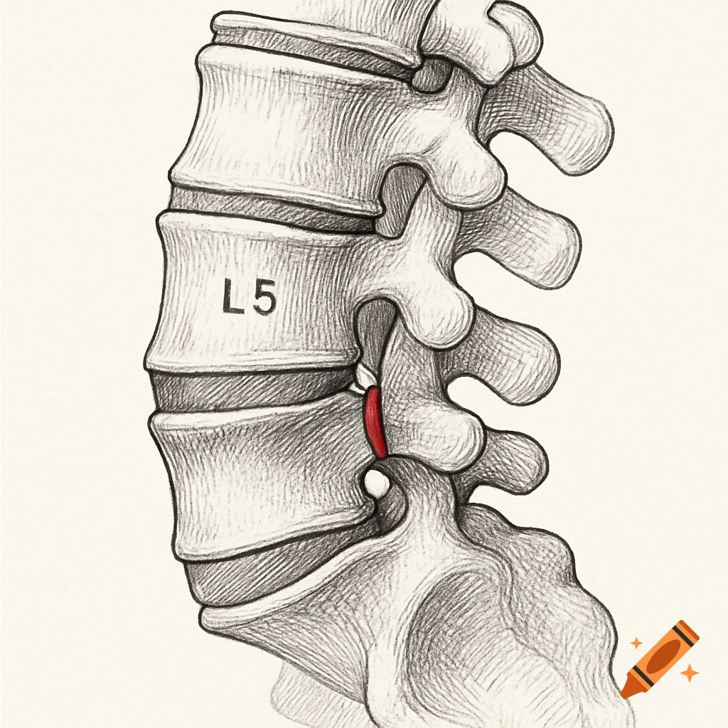
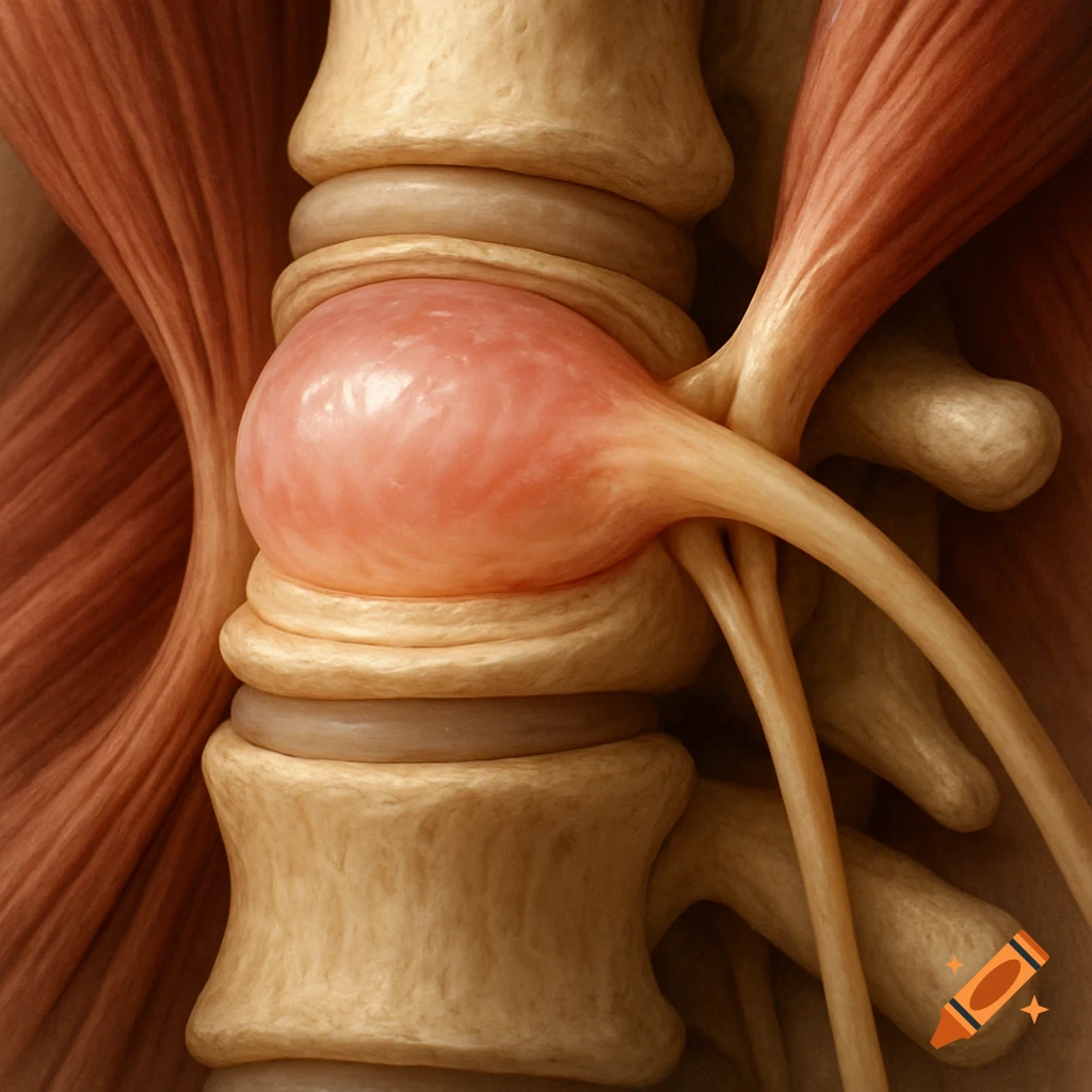
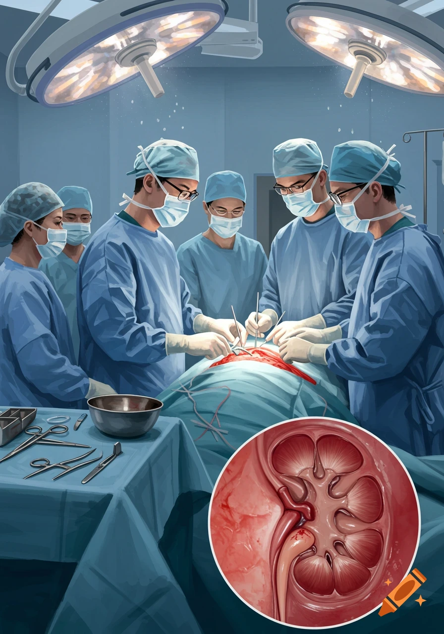
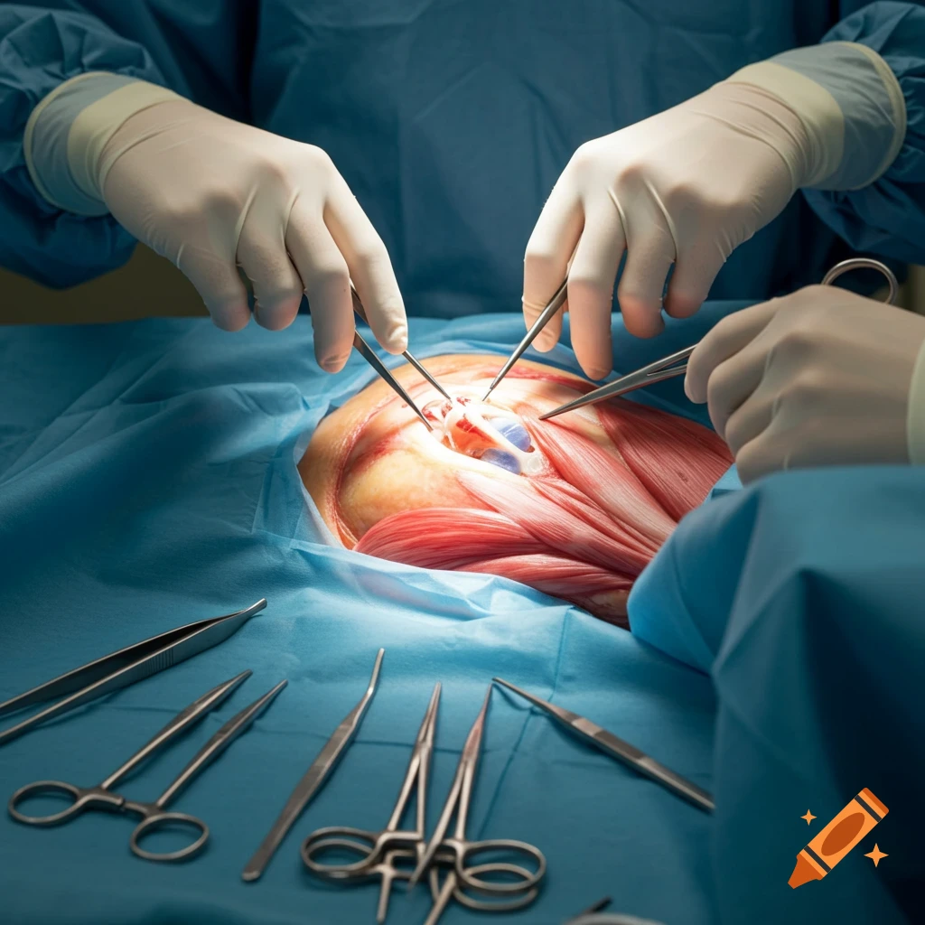
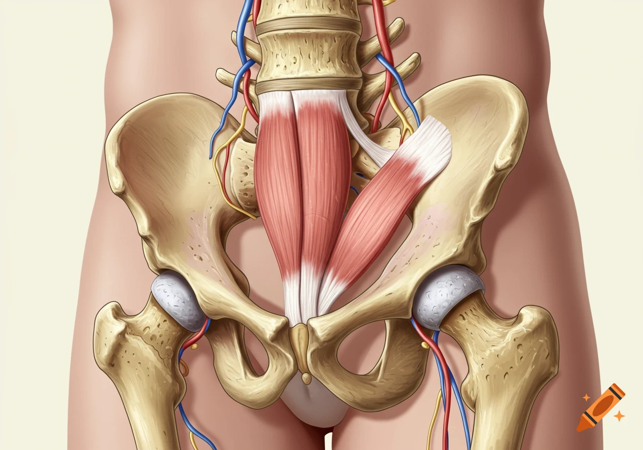
Print me a picture of this procedure The microscope was brought into the field. Soft tissue was cleared off bony edges of the interlaminar window. Using a burr, the caudal edge of the L4 lamina was removed down to the ligamentum flavum. A partial medial facetectomy was performed in order to visualize the lateral recess. The ligamentum flavum was then penetrated with a microcurette and removed piecemeal. The dura sac, traversing nerve root were identified. Full bilateral decompression was performed. An angled hockey stick elevator was tracked underneath the thecal sac proximally and distally and ensured adequate decompression with no remaining fragments compressing the nerve root or the thecal sac. At this point, we were satisfied with the extent of the decompression, the neural elements were seen to be free. See more