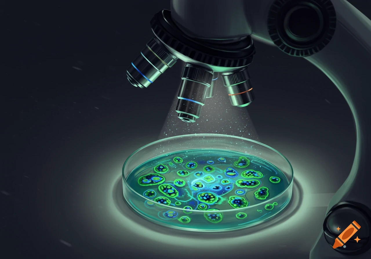
A close-up view of a microscope observing a glowing biological tissue slice on a glass slide. Microscope objectives are visible above.
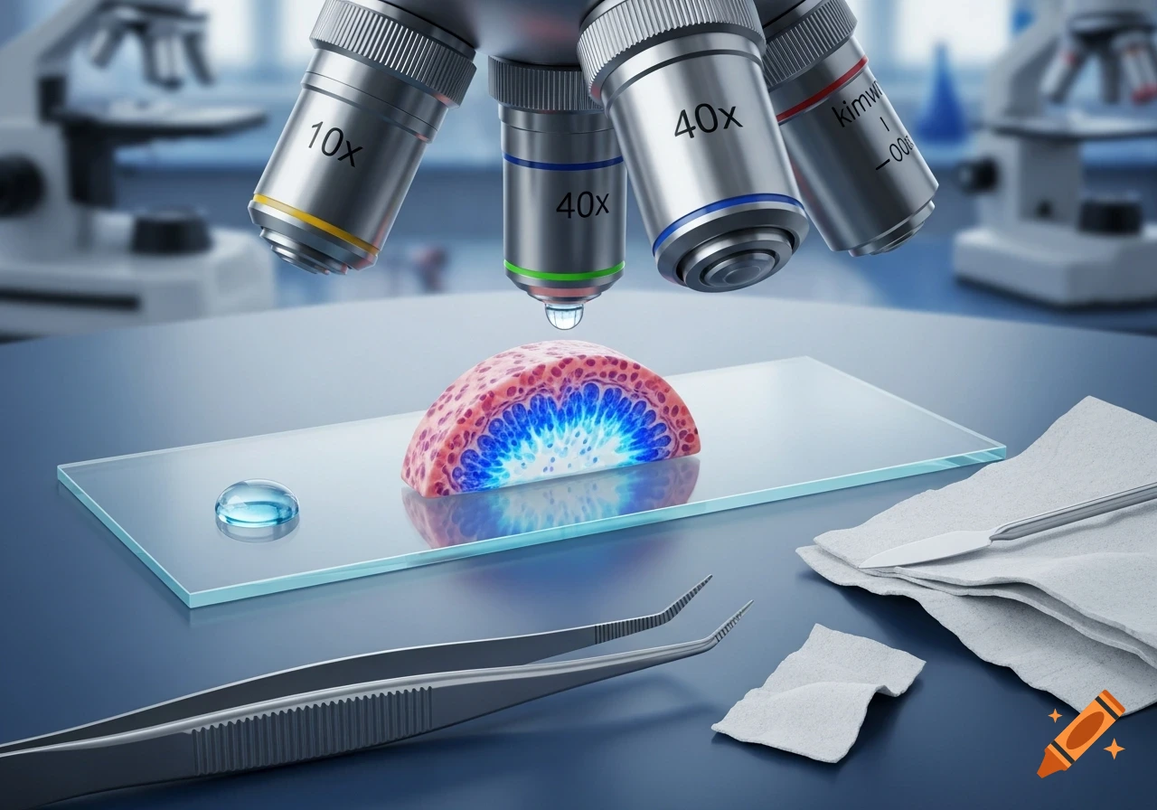
1. Obtain a slide and cover slip, and clean both using a Kimwipe or lens paper. Place a drop of water on the center of the microscope slide. Using tweezers, lay the slice of testicular tissue on top of the drop. Then place a drop of methylene blue stain on top of the slice to help visualize cell structures in the tissue. Place the cover slip over the sample by placing one edge down first and then lowering the cover slip. Examine the sample using a light microscope with the 10x and 40x objective lenses for detailed observation of seminiferous tubules. Sketch your observations below draw as someone not that good at drawing See more