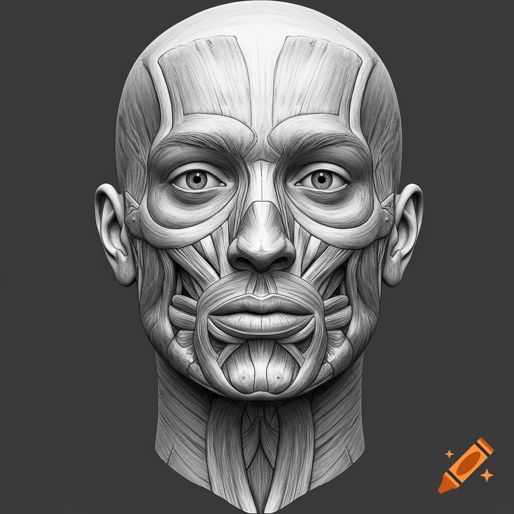
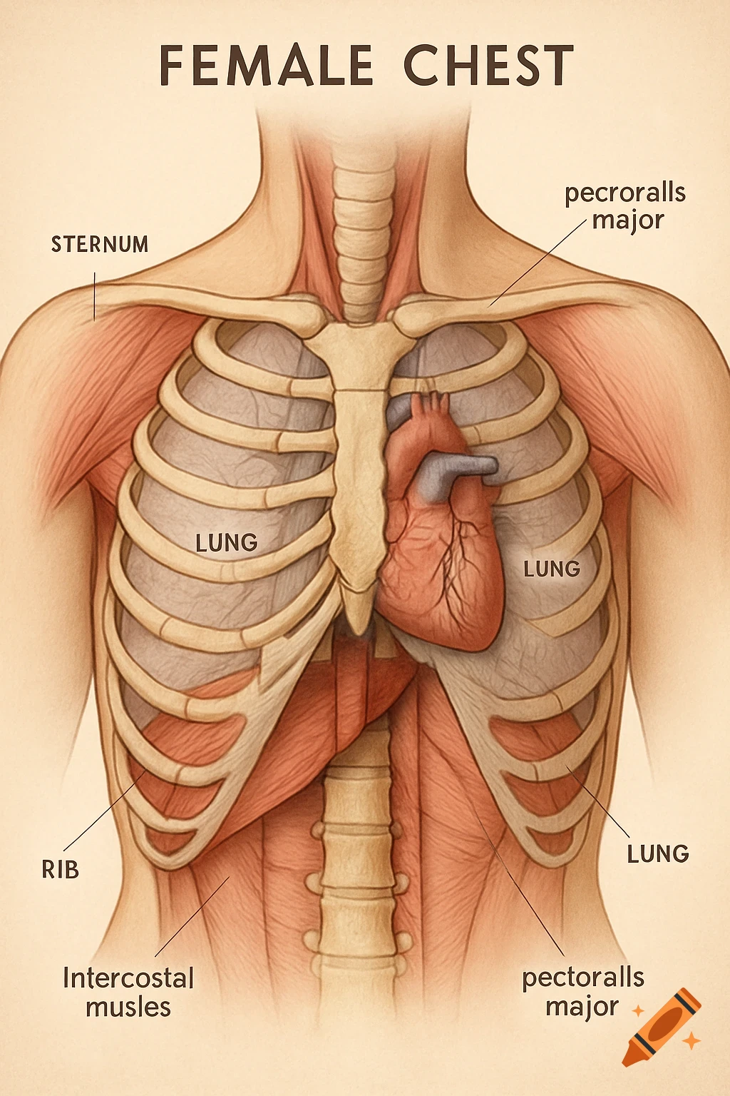
Detailed anatomical illustration of a human head and neck in profile, showing the temporomandibular joint, temporalis, masseter, and sternocleidomastoid muscles with labels.
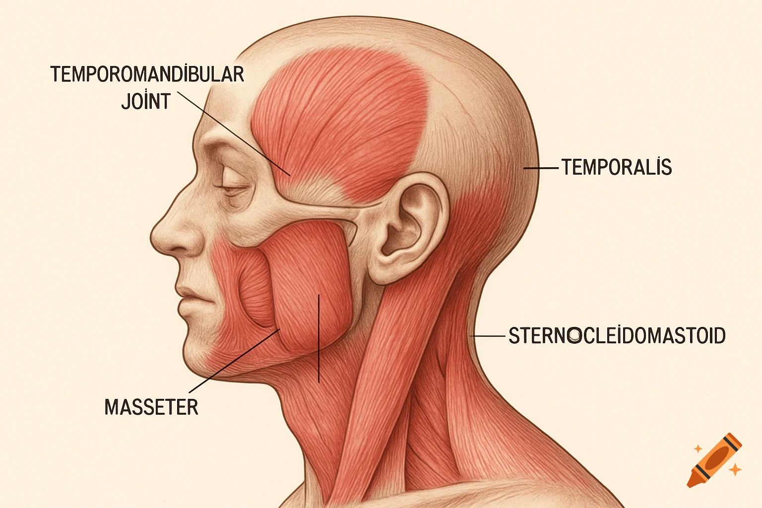
The Temporomandibular Joint (TMJ) would be located just in front of the ear, where the lower jaw connects to the skull. It's often depicted as a small, circular or oval area at this junction. The Masseter muscle is a large, powerful chewing muscle that covers the side of the jaw. It would be shown as a broad muscle extending from the cheekbone down to the angle of the jaw. The Temporalis muscle is a fan-shaped muscle located on the side of the head, covering the temple area. It would originate from the side of the skull and insert into the coronoid process of the mandible (lower jaw). The Sternocleidomastoid (SCM) muscle is a prominent neck muscle. It would run diagonally from behind the ear (mastoid process) down to the collarbone and sternum at the front of the neck. See more