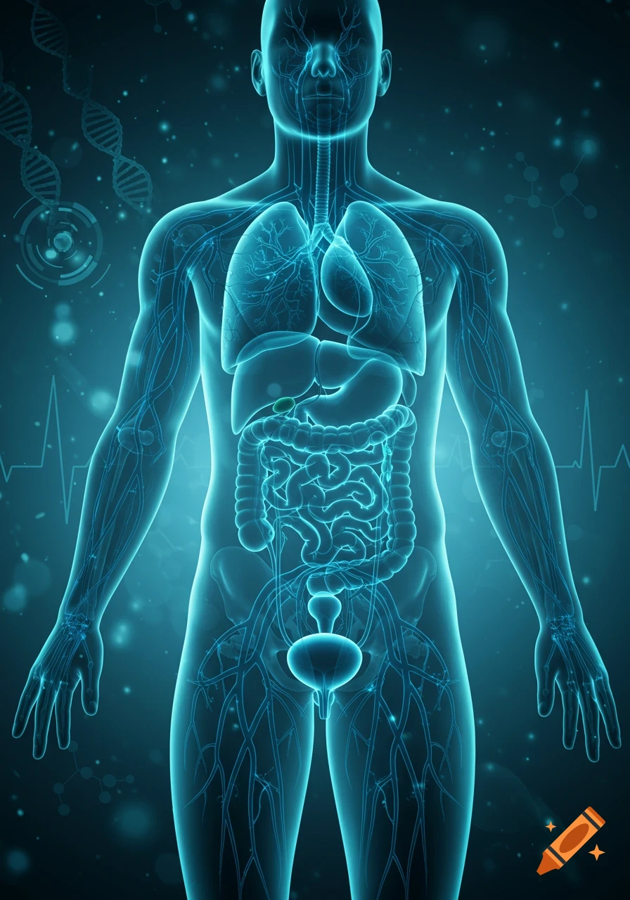
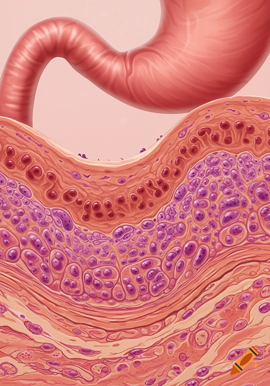
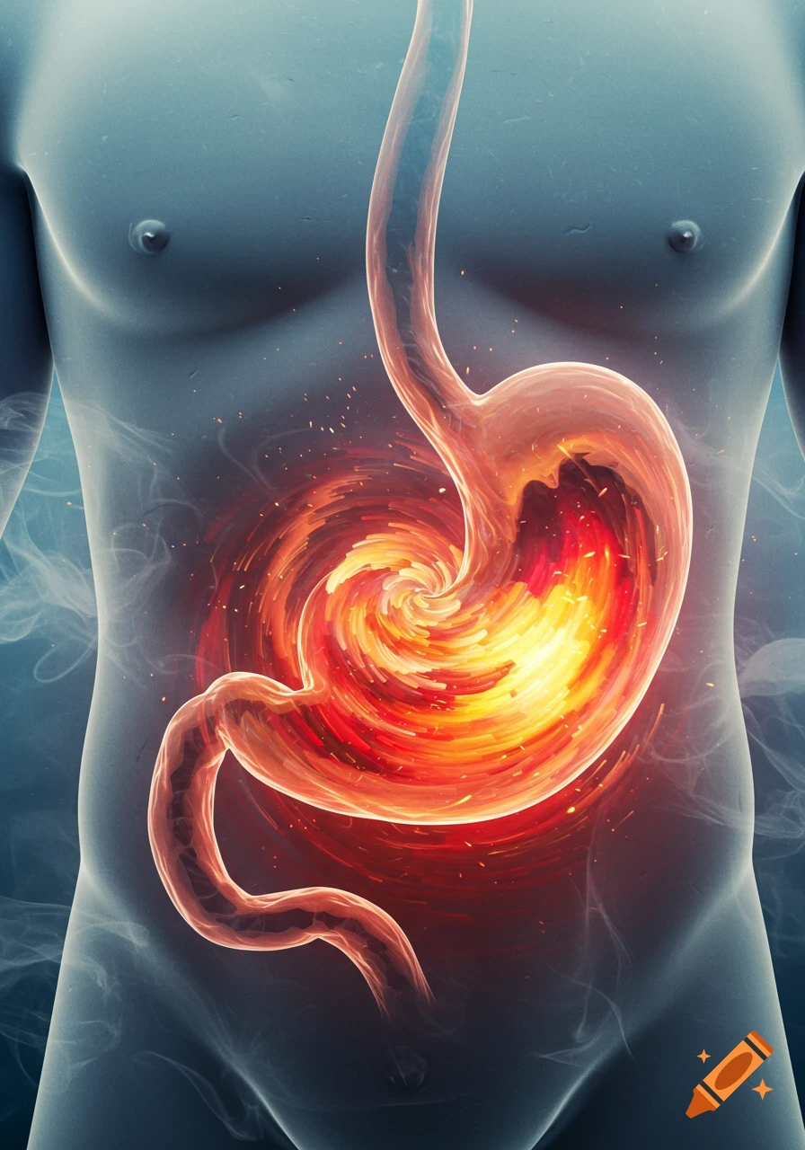
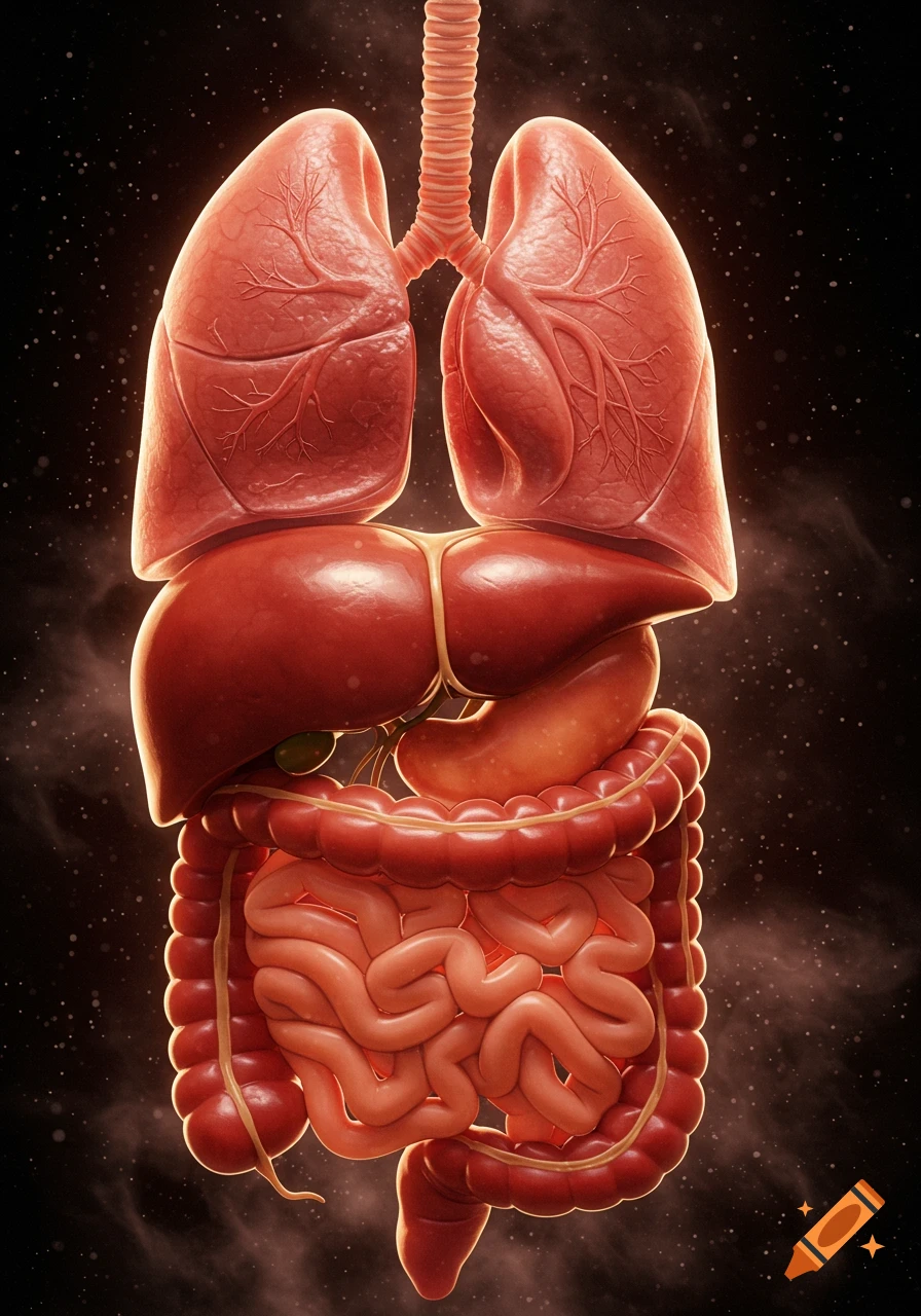
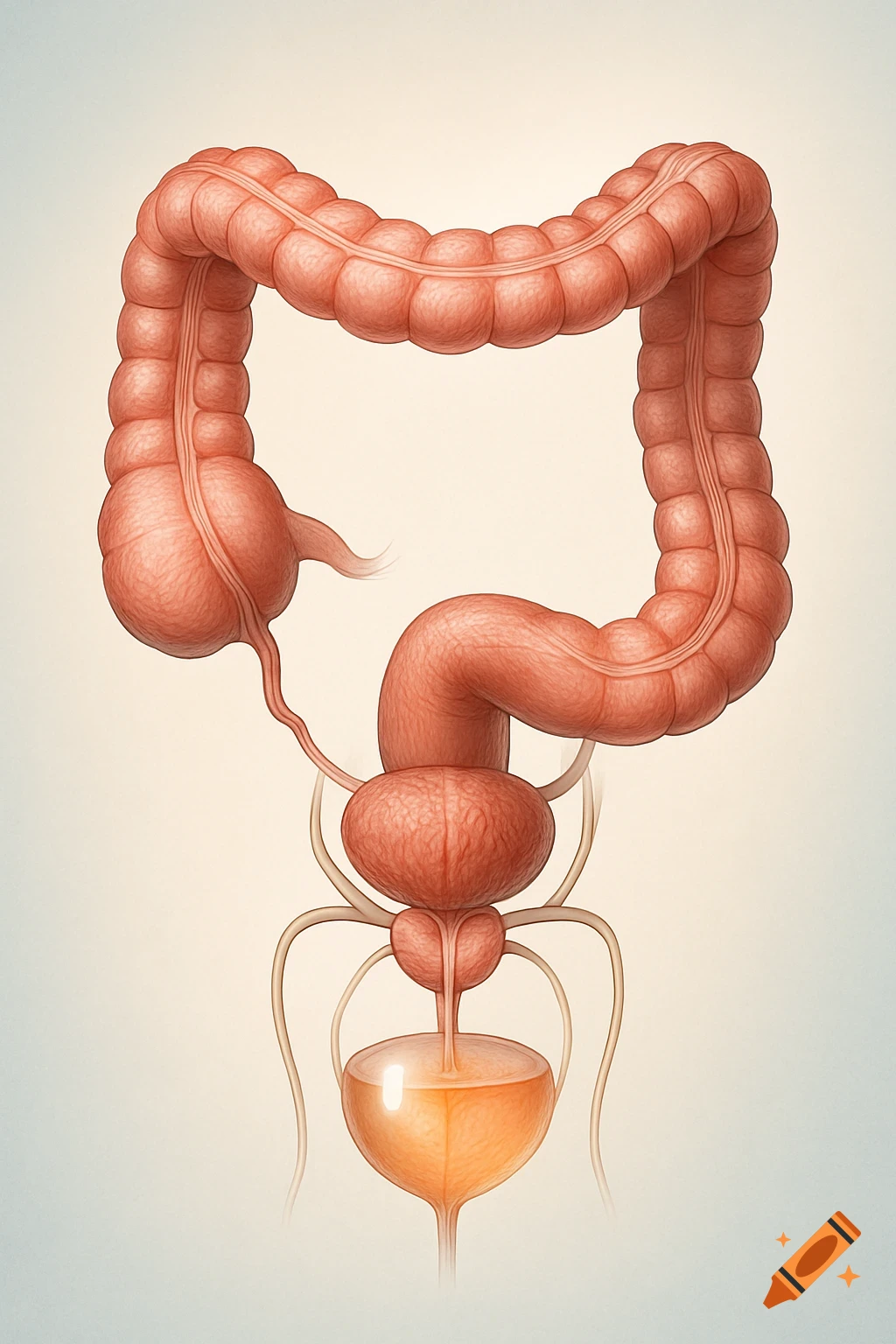
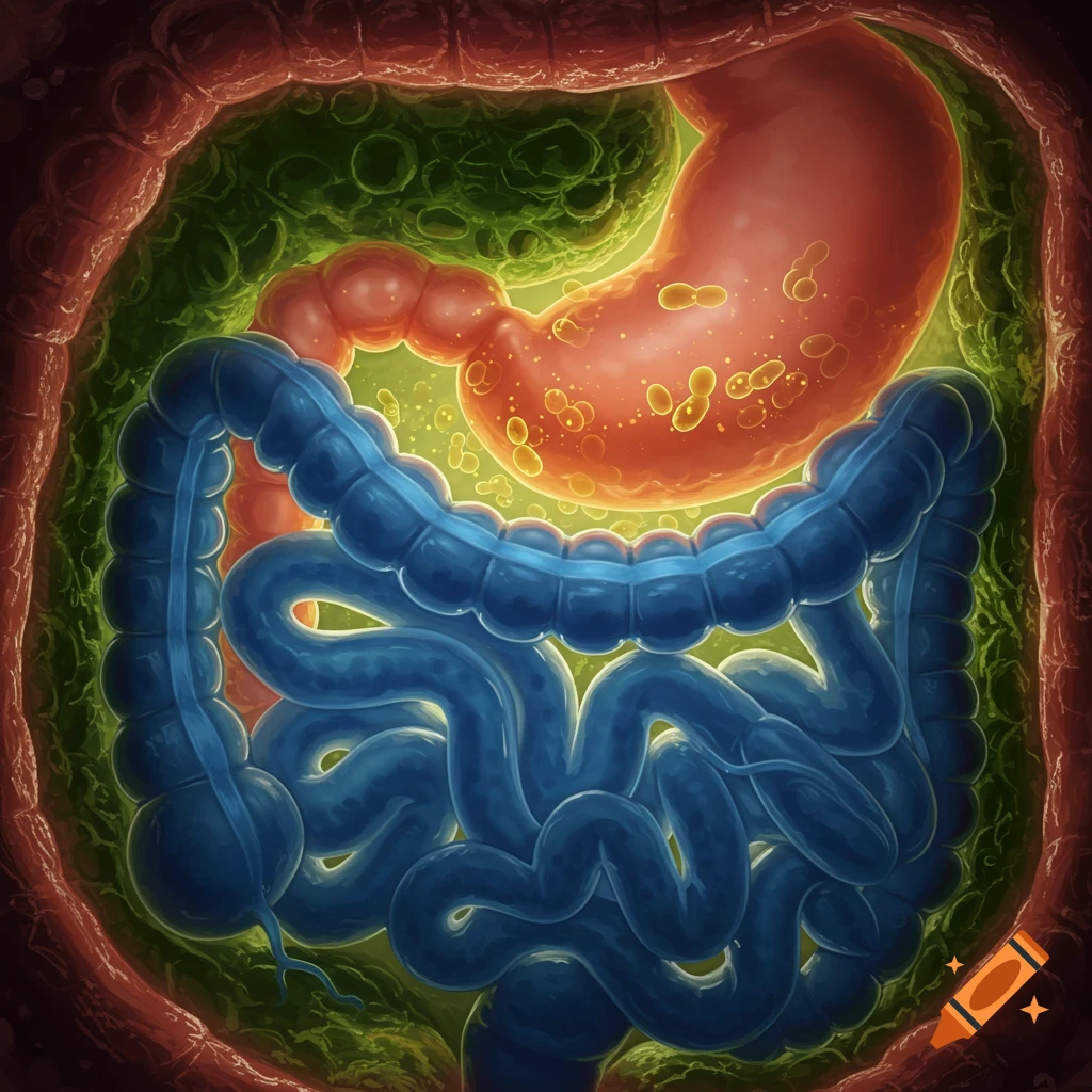
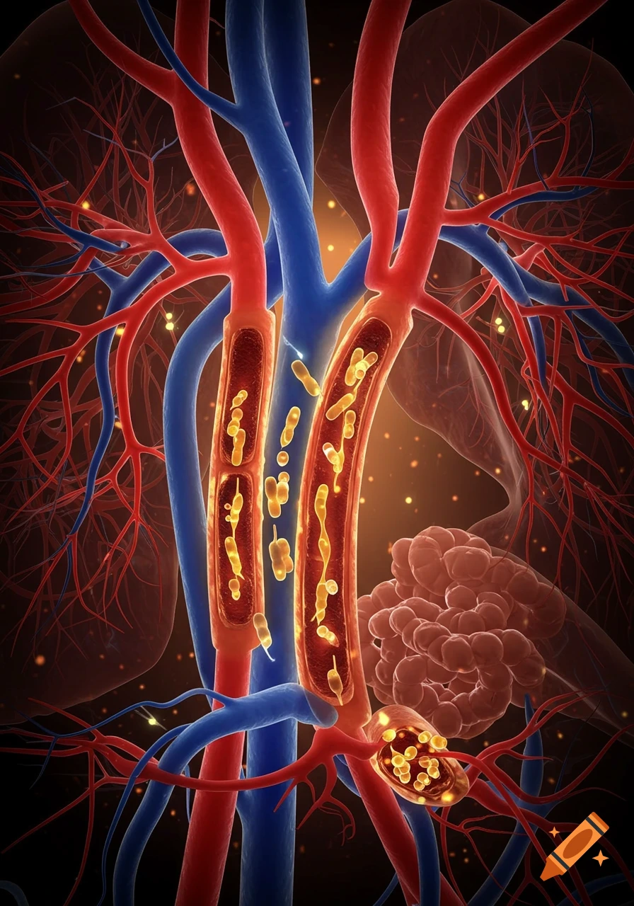
Detailed medical diagram of intestinal loops with magnetic balls causing compression, ischemia, and necrosis in a diagrammatic style.
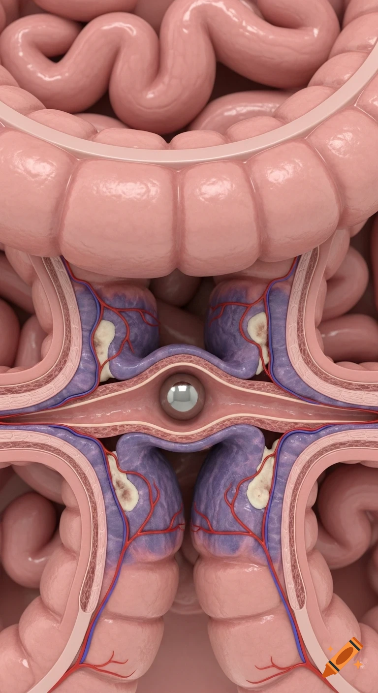
Generate image: Anatomical medical image in a realistic semi-diagrammatic style (like in an educational atlas). Vertical frame (9:16). Scene: two loops of small intestine, each containing a small magnetic ball. The balls are attracted through the intestinal walls, pinching sections of the wall together and causing compression, ischemia, and early signs of necrosis. Style: Accurate medical visualization, without dramatization. Basic requirements for composition and content Two adjacent loops of small intestine are depicted partially dissected/semi-unfolded to reveal the walls (semi-diagrammatic view - outer serous surface + translucent section showing the wall layers). Inside each loop is a small round magnetic ball (shiny metal balls ~3–6 mm). The balls are located in different sections of the loop (not at the same point), but are attracted to each other by gravity through the thin intestinal wall. The following is clearly visible between the spheres: Two intestinal loops pressed against each other; A section of the intestinal wall is compressed, flattened, and thinned; Compression of the mucosa and submucosa; Reduced blood supply—color change, visible compressed vessels. Color differentiation: Normal intestinal tissue—pink (soft pink tones); The area of compression/compression—dark purple-bluish (with signs of ischemia); Small whitish areas in the compression zone—the beginning of necrosis; Vessels—a fine network of blood vessels in the wall; in the compression zone, they See more